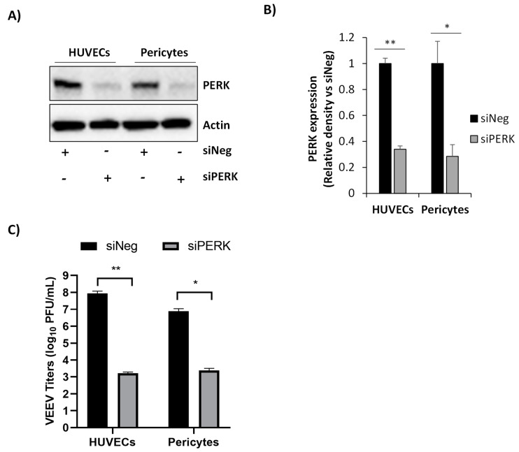
Figure 3.
Loss of PERK decreases VEEV titers in human primary pericytes and human umbilical vein endothelial cells (HUVECs). (A) Pericytes or HUVECs were transfected with 100 nM of siNeg or siPERK siRNAs. Cell lysates were collected 48 h post transfection and analyzed by immunoblot. PVDF membranes were probed for levels of PERK. β-actin was used as a loading control. (B) Quantitative data of panel A. PERK protein levels were normalized to β-actin and normalized values were calculated relative to siNeg-transfected cells. (C) At 48 h post transfection, cells were infected with VEEV TC-83 (MOI 5), and viral replication was analyzed using supernatants collected at 18 hpi via plaque assays in Vero cells. Data are expressed as the mean ± SD (n = 3). * p ≤ 0.05, ** p ≤ 0.01.

