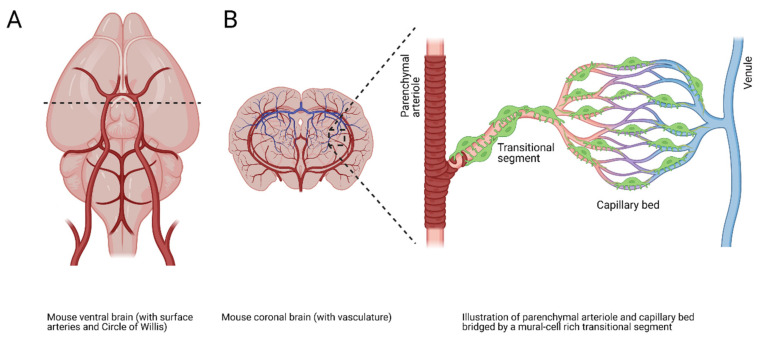
Figure 4.
Cerebral vascular overview and microvascular schematic. (A) Illustration of the mouse ventral brain with visible surface vasculature. Dotted line indicates coronal section in (B). (B) Illustration of a mouse coronal brain section showing a complex vascular network composed of a parenchymal arteriole, a mural-cell–ensheathed transitional segment, a capillary bed with pericytes, and an exiting venule.

