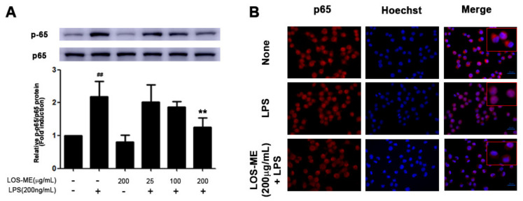
Figure 4.
Effect of LOS-ME on NF-κB p65 phosphorylation in LPS-activated BV2 cells. BV2 microglia cells were treated with LOS-ME (200 μg/mL) and LPS (200 ng/mL) for 30 min and confirmed by Western blot. (A) protein expression of p65 and p-p65 with corresponding fold change. (B) BV2 microglia cells were treated with LOS-ME (200 µg/mL) and LPS (200 ng/mL) for 30 min and confirmed by ICC assay. Translocation of p65 protein was determined using an anti-p65 antibody and Alexa Fluor 568-labeled goat anti-mice antibody on immunofluorescence. Data are presented as mean ± SD (n = 3). ## p < 0.01 compared with control group, ** p < 0.01 compared with LPS group by one-way ANOVA.

