Figure 3.
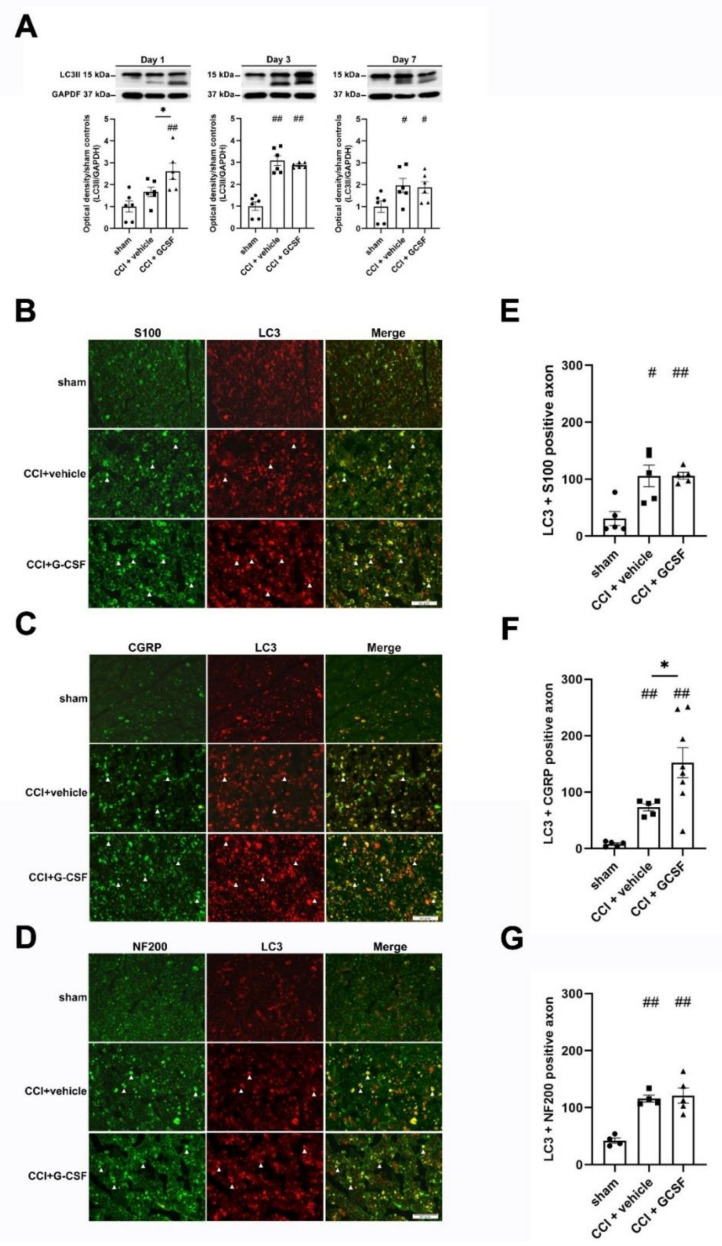
G-CSF upregulated autophagy in the injured sciatic nerve on the 1st day after nerve injury. (A) Western blot analysis revealed significantly higher LC3II protein levels in the injured sciatic nerve in G-CSF-treated CCI rats than in vehicle-treated CCI rats and sham control rats on the 1st day after nerve injury (* p < 0.05, ## p < 0.01: G-CSF-treated CCI rats compared to vehicle-treated CCI rats and sham control rats). However, there was no significant difference in LC3II protein levels in vehicle-treated CCI rats and sham control rats on the 1st day after nerve injury. LC3II protein levels in the injured sciatic nerve were significantly higher in vehicle- and G-CSF-treated CCI rats than in sham control rats from the 3rd to the 7th day after nerve injury. (## p < 0.01, # p < 0.05: the G-CSF-treated and vehicle-treated groups compared to the sham controls on the 3rd and 7th days after nerve injury). However, there was no significant difference between vehicle-treated CCI rats and GCSF-treated CCI rats. The data are shown as the means ± SEM. n = 6 in each group. (B–D) Representative images showing LC3-positive fibers (white arrowhead) in the injured sciatic nerve in sham control rats, vehicle-treated CCI rats and G-CSF-treated CCI rats on the 1st day after nerve injury. LC3-positive fibers were costained with S100 (a marker of the β-subunit of Schwann cells), NF200 (a marker of large-diameter myelinated axons) and CGRP (a marker of small- to medium-diameter myelinated and unmyelinated peptidergic axons). Scale bars = 50 µm. (E–G) Significantly higher numbers of axons that were positive for LC3 + S100 were observed in G-CSF-treated CCI rats and vehicle-treated CCI rats than in sham control rats on the 1st day after nerve injury. (## p < 0.01, # p < 0.05: the G-CSF-treated CCI and vehicle-treated CCI groups compared to the sham controls). However, there was no significant difference between GCSF-treated CCI rats and vehicle-treated CCI rats. Significantly higher numbers of axons that were positive for LC3 + NF200 were also observed in G-CSF-treated CCI rats and vehicle-treated CCI rats than in sham control rats on the 1st day after nerve injury. (## p < 0.01, ## p < 0.01: the G-CSF-treated CCI and vehicle-treated CCI groups compared to the sham controls). However, there was no significant difference between GCSF-treated CCI rats and vehicle-treated CCI rats. G-CSF-treated CCI rats showed significantly higher numbers of LC3 + CGRP-positive axons than sham control rats and vehicle-treated CCI rats on the 1st day after nerve injury (* p < 0.05: the G-CSF-treated CCI group compared to the vehicle-treated CCI group; ## p < 0.01: the G-CSF-treated CCI group compared to the sham controls). There were also significantly higher numbers of LC3 + CGRP-positive axons in the vehicle-treated CCI group than in the sham controls. (## p < 0.01: the vehicle-treated CCI group compared to the sham controls). The data are shown as the means ± SEM. n = 5 in each group.

