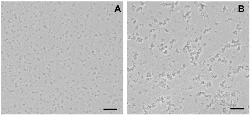
Figure 5.
Light microscopy images of rat liver mitochondria incubated with F16-betulin conjugate. Panel (A) shows control sample (without additions). Panel (B) shows a sample of mitochondria captured after incubation with 30 μM conjugate. Typical images are presented in two sets for each experimental condition. Scale bar is 10 μm.

