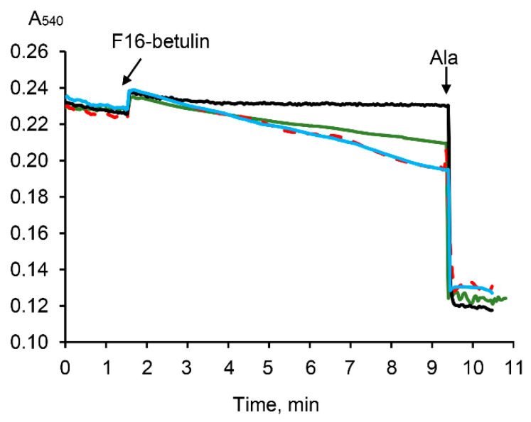
Figure 6.
Effect of F16-betulin conjugate on the optical density of a suspension of liver mitochondria. Additions: 30 µM of F16-betulin conjugate in the presence of glutamate plus malate (green line) and succinate plus rotenone (blue line). The effect of 1 μM cyclosporin A (CsA) on changes in the optical density of succinate-fueled mitochondria induced by the conjugate is shown by the red dashed curve. Black line reflects the kinetics of changes in the optical density in the absence of oxidation substrates. Alamethicin (Ala, 5 μg/mL) was used to show the maximum possible decrease in the optical density of the mitochondrial suspension at the end of the recordings. A similar pattern was observed in three independent experiments.

