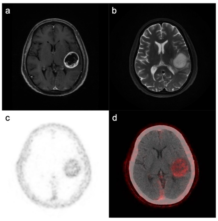
Figure 2.
Magnetic resonance imaging (MRI) and FAPI PET/CT in GBM. An IDH-wildtype GBM from a 71-year-old woman displayed ring-like contrast enhancement with hypointense T1-weighted and hyperintense T2-weighted signals (a,b) from central tissue. 68Ga-FAPI PET and merged PET/CT images (c,d) showed elevated radioactivity in the whole tumor area (including ring-like contrast enhancement and noncontrast-enhanced tissue).

