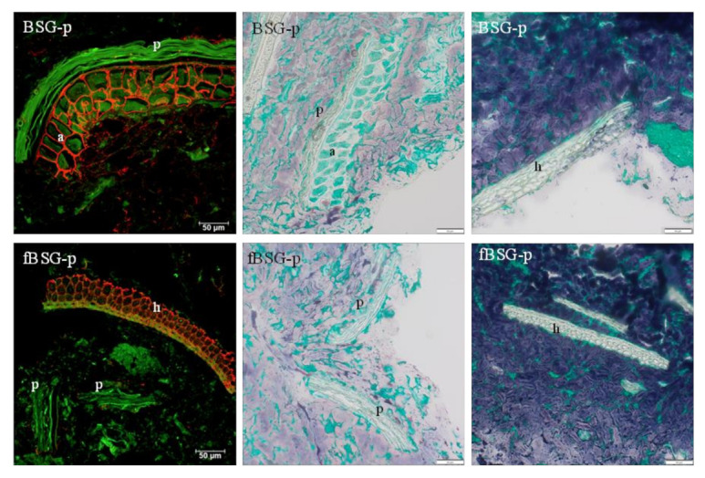
Figure 3.

CLSM (left) and brightfield (middle, right) micrographs of pasta containing fermented and native brewers’ spent grain: BSG-p, pasta containing native brewers’ spent grain; fBSG-p, pasta containing fermented brewers’ spent grain. In CLSM micrographs arabinoxylan is shown in red and autofluorescence (cell walls, protein) is shown in green. Bright field micrographs show starch in blue and protein in green. h= hull (palea and lemma), a = aleurone, p = pericarp. Scale bars 50 µm.
