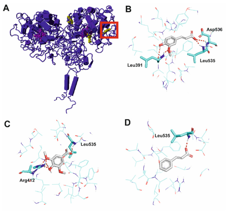
Figure 2.
Molecular interactions of activators with thyroid peroxidase (TPO). (A) Ferulic acid bound to three considered binding pockets. The protein is shown in dark blue cartoon representation, ferulic acid in yellow ball representation and heme in magenta ball representation. The selected pocket is marked with a red square. (B) Ferulic acid, (C) syringic acid and (D) trans-cinnamic acid in a binding pocket of TPO. The protein is shown with cyan carbon atoms in wire representation with the most important residues shown as sticks. Activators depicted with gray carbon atoms in stick representation. Polar bonds are shown as red dashes. Nonpolar hydrogen atoms omitted for clarity.

