
Figure 7. Multinuclear cell fusion rate with expression of myogenesis markers in 2-week differentiation.
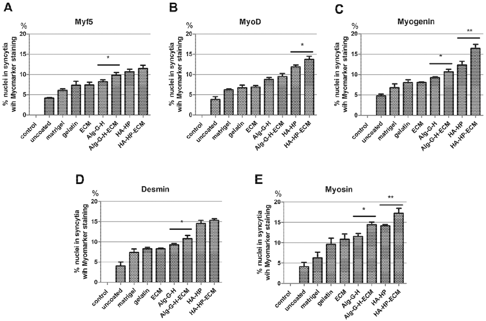
Percentage of nuclei in syncytia with positive staining for A) MyoD, B) Myf5, C) Myogenin, D) Desmin, and E) Myosin versus overall nuclei assessed, in different coating conditions such as Matrigel®, gelatin, ECM, Alg-G-H, Alg-G-H-ECM, HA-HP, HA-HP-ECM, compared with uncoated conditions and normal hSMPCs as control. Histograms are means ± SEM of four experiments on each independent culture condition. Data were analyzed by the one-way analysis of variance (ANOVA) for all groups, and then Student’s t-test for comparison of two groups. Results are presented as mean ± SEM, * p<0.05, ** p<0.01.