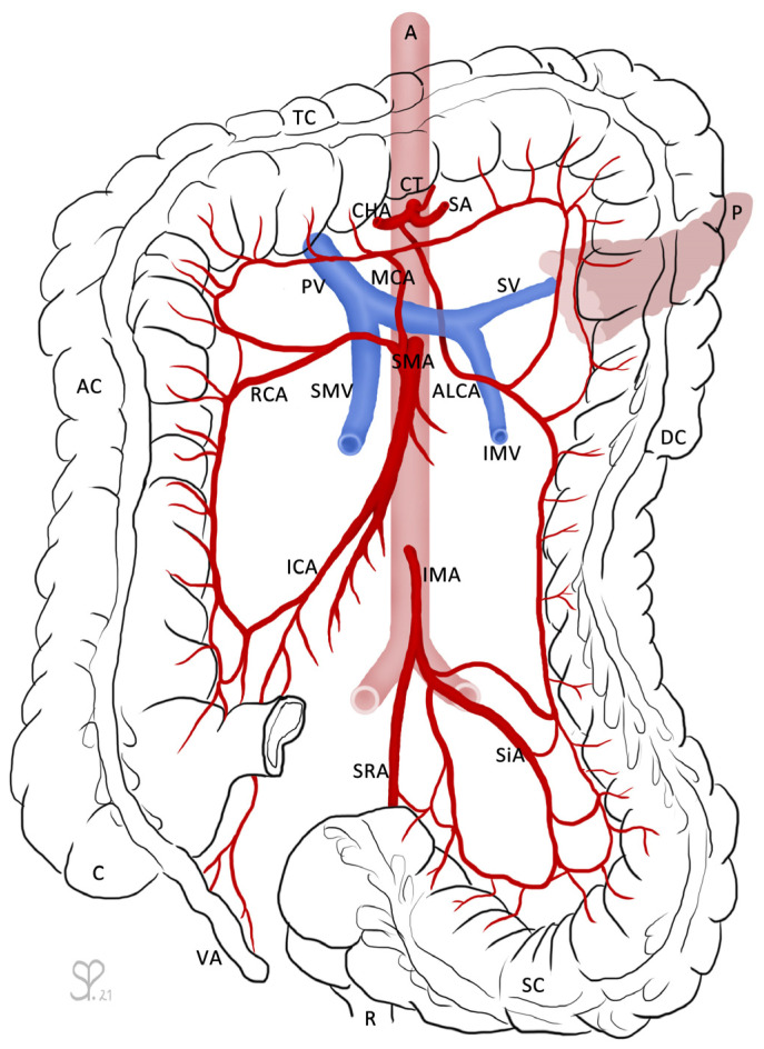
Figure 1.
The large intestines (caecum (C) with vermiform appendix (VA), ascending colon (AC), transverse colon (TC), descending colon (DC), sigmoid colon (SC)), as well as the rectum (R) and pancreas (P) are outlined for orientation. From the abdominal aorta (A) arises the coeliac trunk (CT), which branches into the common hepatic artery (CHA), the splenic artery (SA), and the left gastric artery (not labeled). The aberrant left colic artery (ALCA) descends shortly after the branching from the CHA and runs underneath the pancreas and splenic vein (SV). The SV receives the inferior mesenteric vein (IMV) and fuses with the superior mesenteric vein (SMV) to become the portal vein (PV). The superior mesenteric artery (SMA) branches into the middle colic artery (MCA), right colic artery (RCA), and ileocolic artery (ICA). The inferior mesenteric artery (IMA) divides into the sigmoid arteries (SiA) and superior rectal artery (SRA). The ALCA forms anastomoses with the MCA via its ascending branch and with the SiA via its descending branch. Drawing by Sandra Petzold.

