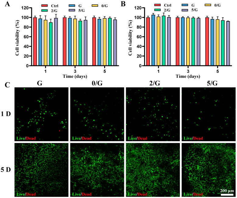
Figure 3.
The cytocompatibility of hydrogels. (A) Proliferation of HUVECs cultured in different hydrogel extracts (n = 6). (B) Proliferation of L929 cells cultured in different hydrogel extracts (n = 6). (C) Live/dead staining images at 1 and 5 days after L929 cells encapsulation in different hydrogels.

