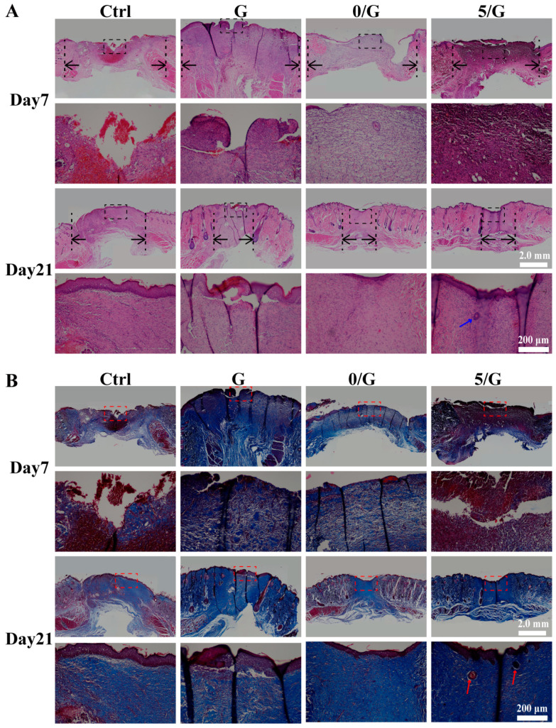
Figure 7.
Histology analysis in wounds treated with different hydrogels. (A) H&E staining of wound tissue after 7 and 21 days with different treatments. Black arrows indicated microscopic wound edges, blue arrow indicates skin appendages. (B) Masson’s trichrome staining of wound tissue after 7 and 21 days with different treatments, red arrow indicates skin appendages.

