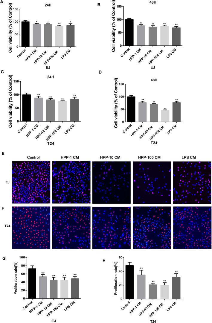
Fig. 2.
Cell viability and proliferation were restrained when incubated with the aforementioned supernatant. a-d Change in the cell viability (% of the control group) when human bladder cancer cells T24 and EJ were cultured with the supernatant for 24 h or 48 h. (e-h) Effect of activated macrophages on T24 and EJ cell proliferation observed using EdU staining (200×). Red fluorescence denotes EdU staining, representing proliferating cells. Blue fluorescence denotes DAPI staining, representing the staining of nuclei. Statistical data analysis showed a downward trend in cell proliferation. *P < 0.05; **P < 0.01 compared with the control group(n = 3)

