Figure 2.
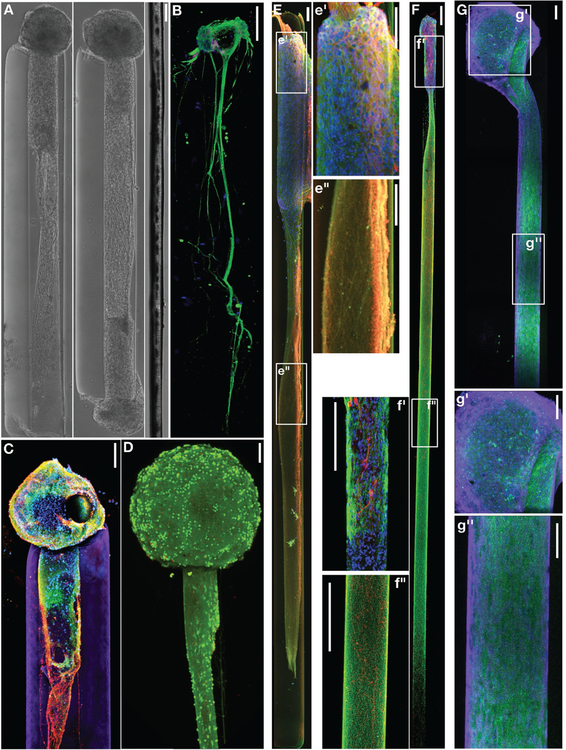
Neuronal-Axonal Living Electrodes: (A) Phase contrast images of unidirectional (left) and bidirectional (middle) “living electrodes” built using cerebral cortical neurons, each at 5 days in vitro (DIV), next to a single human hair (right). (B) Confocal reconstruction of a living electrode built using dorsal root ganglia neurons showing unidirectional axonal tracts immunolabeled to denote neuronal somata (MAP-2; purple) and axons (tau; green), with nuclear counterstain (blue). (C) Confocal reconstruction of a unidirectional, cerebral cortical neuronal living electrode at 11 DIV, immunolabeled for axons (β-tubulin-III; red) and synapses (synapsin; green), with a nuclear counterstain (Hoechst; blue). The surrounding hydrogel micro-column is shown in purple. (D) Confocal reconstruction of a unidirectional cortical neuronal living electrode stained for viability at 10 DIV (green: live cells via calcein-AM; red: nuclei of dead cells via ethidium homodimer-1). Scale bars A-D: 100 μm. (E-G) Long-projecting unidirectional axon-based living electrodes for tailored neuromodulation. (E) Confocal reconstruction of an excitatory living electrode built using neurons derived from the cerebral cortex (predominantly glutamatergic), immunolabeled at 28 DIV for axons (β-tubulin-III; red) and neuronal somata/dendrites (MAP-2; green), with nuclear counterstain (Hoechst; blue). Insets of the aggregate (e’) and axonal (e”) regions are outlined and shown to the right. Scale bars: 100 μm. (F) Confocal reconstruction of a dopaminergic living electrode built using neurons isolated from the ventral mesencephalon (enriched in dopaminergic neurons), immunolabeled at 28 DIV for axons (β-tubulin-III; green) and tyrosine hydroxylase (dopaminergic neurons/axons; red), with nuclear counterstain (Hoechst; blue). Insets of the aggregate (f’) and axonal (f”) regions are outlined and shown to the left. Scale bars: 250 μm. (G) Confocal reconstruction of an inhibitory living electrode built using neurons isolated from the medial ganglionic eminence (source of GABAergic neurons), immunolabeled at 14 DIV for axons (β-tubulin-III; purple) and GABA (inhibitory neurons/axons; green), with nuclear counterstain (Hoechst; blue). Insets of the aggregate (g’) and axonal (g”) regions are outlined and shown below. Scale bars: 100 μm.

