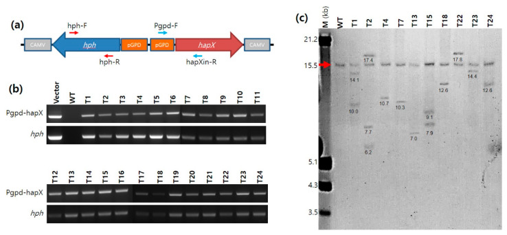
Figure 1.
The transformation of Agaricus bisporus. (a) Gene arrangement of the integrating unit in pBGgHg-hapX. Red arrows and blue arrows indicate the positions of primers for the detection of hph and pGPD-hapX, respectively. CAMVs are CAMV poly(A) signals residing in front LB and RB of pBGgHg-hapX. (b) PCR analysis of the transformants. The amplicons were approximately 500 bp for both targets. (c) Southern blot analysis of the transformants. Genomic DNA (20 μg) was isolated from liquid cultures, digested with AgeI and SacI, and probed with an ~675 bp DIG-labeled hapX gene sequence. Lanes; M, DNA molecular size marker (kb); WT, untransformed A. bisporus; T1 to T24, transformants. The red arrow indicates the original hapX gene fragment. Numbers under the DNA bands indicate the sizes of genomic DNA fragments.

