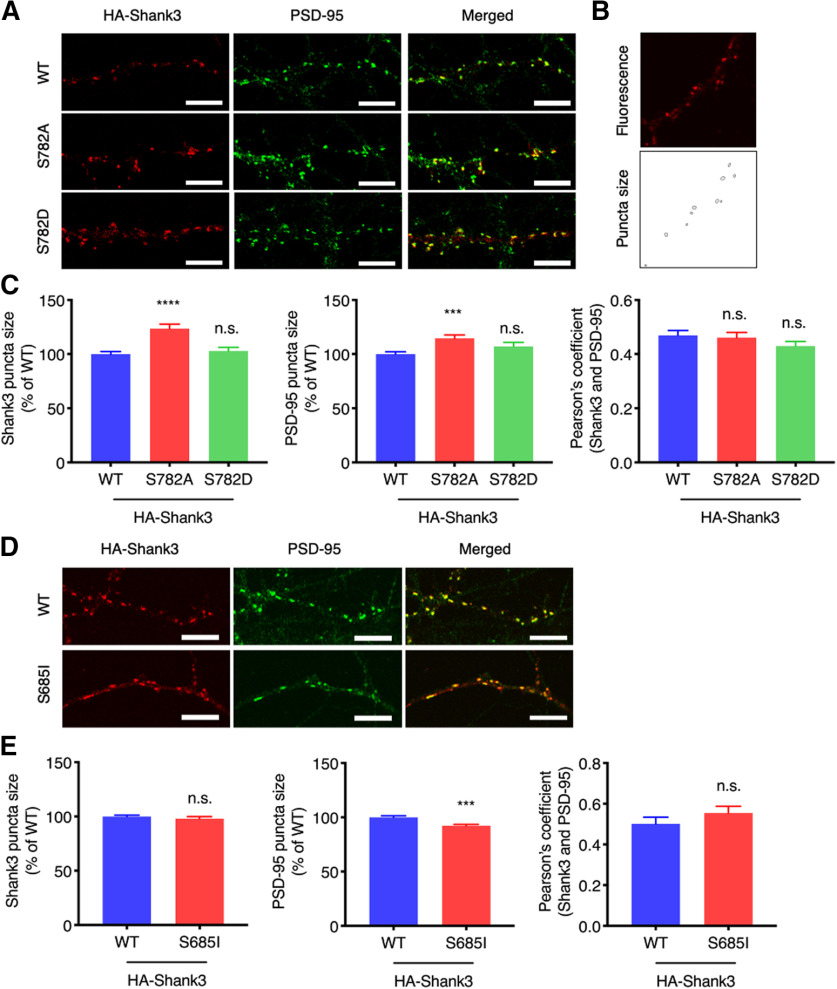
Figure 5.
Shank3 S782 phosphorylation affects Shank3 enrichment and synapse size. A, HA-Shank3 (WT, S782A, or S782D) was expressed in cultured rat hippocampal neurons. HA-Shank3 was stained with anti-HA and Alexa Fluor 555-conjugated secondary antibody (red). Endogenous PSD-95 was labeled with anti-PSD-95 antibody and Alexa Fluor 488-conjugated secondary antibody (green). Region from the secondary dendrites is shown. Scale bar: 5 μm. B, Fluorescence signal was adjusted to measure perimeter of synaptic puncta. C, Region from the secondary dendrites were analyzed for puncta size and Pearson’s coefficient. Graph indicates mean ± SEM (n = 28 for WT, n = 26 for S782A and S782D); ****p < 0.0001 and ***p < 0.0002 using one-way ANOVA with Dunnett’s multiple comparison test. Colocalization analysis between endogenous Shank3 and PSD-95 on CaMKII pharmacological activation in neurons are presented in Extended Data Figure 5-1. D, HA-Shank3 (WT or S685I) was expressed in cultured rat hippocampal neurons. HA-Shank3 and endogenous PSD-95 were stained and analyzed as above. Region from the secondary dendrites is shown. Scale bar: 5 μm. E, Graph indicates mean ± SEM (n = 24 for WT, n = 30 for S685I); ***p < 0.0002 using an unpaired t test. n.s., not significant.

