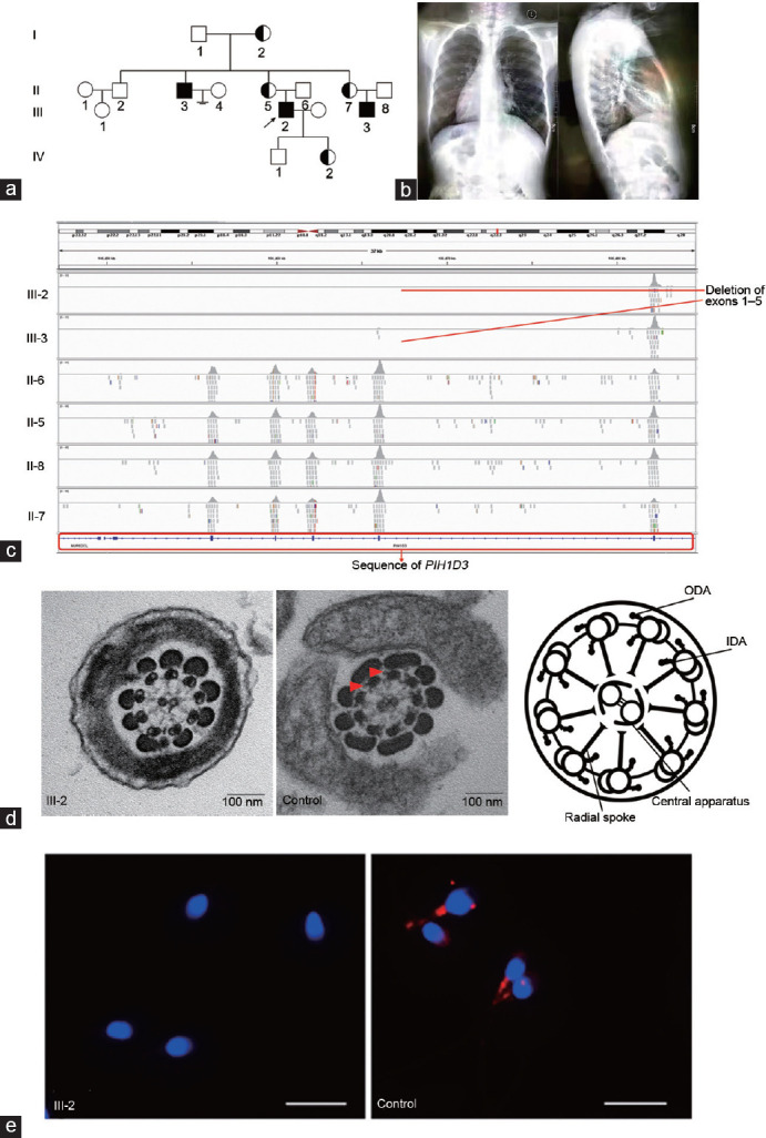
Figure 1.

Results of the mutational analyses in PIH1D3. Pedigrees of the PCD-affected family and clinical features. (a) Hemizygous loss-of-function mutations in PIH1D3 located on the X chromosome were identified in one family. Pedigree of the PCD-affected family. PCD-affected siblings are indicated in black, and the unaffected siblings are indicated in white. (b) Chest X-ray shows situs inversus totalis, chronic airway disease with bronchiectasis in the middle lobe, and situs inversus totalis in III-3. (c) All blood samples were detected with whole-exome sequencing. The proband and his cousin had deletion of the PIH1D3 gene exons 1–5. (d) Analysis of transmission electron microscopy showing the cross-section arrangement of cilia from a healthy control (middle) and an image (right) of the main structures of the 9 + 2 motile axoneme, including the ODA and IDA. However, cross-sections from the respiratory cilia from III-2 showed a normal 9 + 2 architecture with absence of outer and inner dynein arms (red arrowheads). Scale bars = 100 nm. (e) Sperm of healthy men and III-2 were labeled with anti-PIH1D3 antibodies (red). The proteins are localized in the tails of sperm of controls. In contrast, PIH1D3 expression was absent or severely reduced in sperm from III-2. Scale bars = 10 μm. PIH1D3: PIH1 domain containing 3; PCD: primary ciliary dyskinesia; ODA: outer dynein arms; IDA: inner dynein arms.
