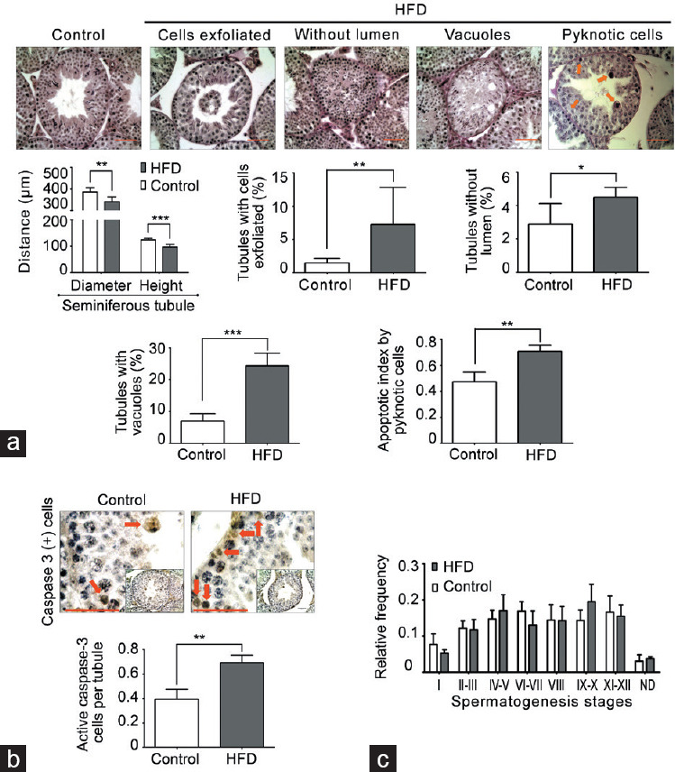
Figure 2.

Degeneration-atrophy of seminiferous tubules and increase in apoptosis in HFD-fed mice. (a) Morphometric analysis of seminiferous tubules (diameter and epithelial height). In addition, images of testicular sections stained with PAS and hematoxylin that present different types of seminiferous tubule degeneration/atrophy and germ cell death by pyknotic cells (orange arrow) found in HFD-fed compared with chow-fed mice. The quantification of histological alterations and germ cell death in HFD-fed versus chow-fed mice is shown at the bottom. Scale bars = 100 μm. (b) Evaluation of germ cell apoptosis by caspase-3-positive cells (red arrow). Scale bars = 100 μm. (c) Frequency of seminiferous epithelial cycles. All graphs represent the mean ± s.d., n = 4. Data were statistically analyzed with an unpaired t-test: *P < 0.05, **P < 0.01, ***P < 0.001. s.d.: standard deviation; HFD: high-fat diet; PAS: periodic acid–Schiff.
