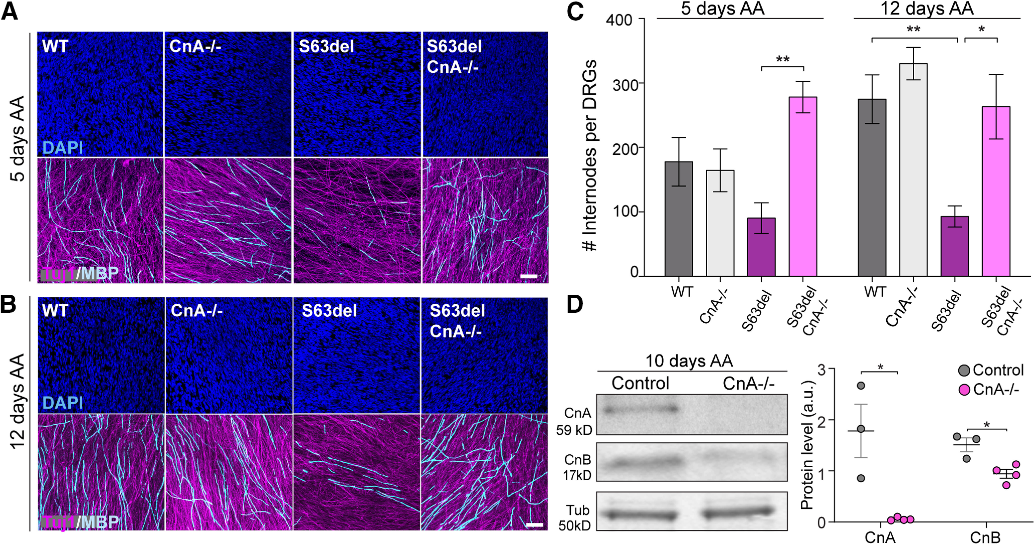
Figure 6.

Ablation of CnA in DRG explants improves S63del hypomyelination in vitro. DRGs were dissected from E13.5 embryos, and myelination was induced with 50 μg/ml of AA after 5 d in culture. A, B, In vitro myelination was assessed in WT, CnA−/−, S63del, and S63del/CnA−/− DRGs. Myelin was visualized by MBP staining (cyan) on DRGs after 5 (A) and 12 d (B) of AA treatment. Axon and nuclei were also revealed with Tuj-1 (magenta) and Dapi (blue), respectively. Scale bar, 50 μm. C, Graphs represent MBP quantification (A,B). n = 30-108 acquisitions from 5-18 DRGs per genotype. DRGs were isolated from E13.5 embryos from 3 different female mice, in two separate dissections. Error bars indicate SEM. *p < 0.05; **p < 0.01; one-way ANOVA with Bonferroni's multiple comparison. D, Left, WB for CnA and CnB was performed on WT and CnA+/− (Control) and CnA−/− DRGs after 10 d of myelination. β-Tubulin (Tub) was used as loading control. Right, Graph represents the mean of CnA and CnB WB in controls and CnA−/− DRGs. Error bars indicate SEM. *p = 0.01 (Student's t test).
