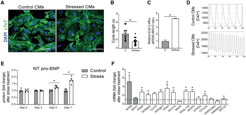
Figure 4.
Genes induced in pathological CMs are increased in stressed human iPS-derived CMs. (A) Representative images of hiPS-CMs in control conditions or after NE and AngII treatment. Immunofluorescence for cardiac troponin T (cTnT) and nuclei DAPI. (B) Calcium transient analysis of the frequency of spontaneous calcium transients and (C) calcium transient analysis of the calcium release of control (C) or NE/Ang II treated (Stress) CMs. (D) Representative spontaneous calcium transients in control (upper panel) and stressed (lower panel) iPS-derived CMs. (E) Human NT-proBNP content in the supernatant of control and NE/AngII (stress) treated CMs determined by ELISA assay (n = 5). (F) Real-time PCR analysis of stress markers and the newly identified genes on NE-AngII treated CMs. Data are expressed as mean fold change ± SEM; *P < 0.05 compared to control (C) with unpaired t-test (n = 3–9) or in a two-way ANOVA.

