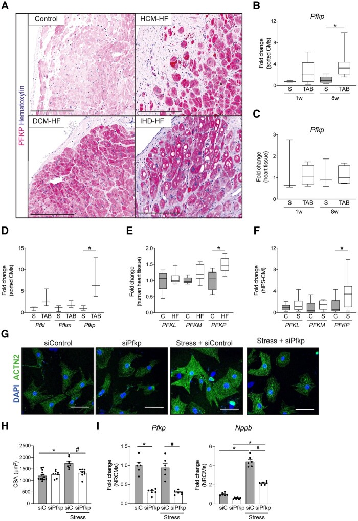
Figure 6.
PFKP is expressed in human failing CMs. (A) Immunohistochemistry of PFKP in healthy and HF dilated cardiomyopathy (DCM-HF), hypertrophic cardiomyopathy (HCM-HF), and ischaemic heart disease (IHD-HF). (B) Real-time PCR of PFKP on mouse sorted CMs (n = 3) on sham (S) or TAB conditions. (C) Real-time PCR of PFKP on mouse heart tissue (n = 4–7) on sham (S) or TAB conditions. (D) Expression analysis of PFK isoforms PFKM (phosphofructokinase-muscle) and PFKL (phosphofructokinase-liver) from bulk RNA sequencing of mouse sorted CMs 8w post TAB (n = 3). (E) Expression analysis of PFK isoforms PFKM (phosphofructokinase-muscle) and PFKL (phosphofructokinase-liver) from RNA sequencing of human failing hearts (n = 5–13). (F) Real-time PCR of PFK- isoforms on NE/AngII treated hiPS-derived CMs (n = 6–9). (G) Representative images of control or PE-treated NRCMs transfected with scramble siRNA control or Pfkp siRNA. Immunofluorescence for sarcomeric α actinin (ACTN2) and nuclei DAPI. (H) Cardiomyocyte cross-sectional area (CSA) quantification (n = >120), and (I) real-time PCR analysis of the cardiac stress markers Nppb and Pfkp of control or PE-treated NRCMs transfected with Pfkp siRNA (siPFKP) or scrambled siRNA control (si-C) (n = 6). (B–F) Data are expressed as average fold change with box (25–75 percentile) and whiskers (min–max); *P < 0.05 in one way ANOVA with Sidak post hoc test. (H and I) Data are expressed as mean fold change ± SEM; *P < 0.05 compared to control (C) or sham (S) and #P < 0.05 compared to siRNA control-stressed (si-C-Stress) in a one-way ANOVA or unpaired t-test.

