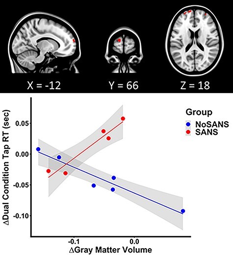
Figure 8 .

Differential GM structure and function association between the SANS and NoSANS groups. The SANS group individuals who showed greater increases in the frontal pole volume from BDC-7 to HDT 29 exhibited greater slowing of dual condition finger tap RTs, while the NoSANS group showed the contrasting brain-behavioral association pattern. The scatter plot depicts the pre-to-post bed rest mean change in GM volume of the peak voxel of the largest left frontal pole cluster (MNI coordinate: −12, 66, 18) as a function of the pre-to-post bed rest change in dual condition finger tap RTs. The shaded areas indicate the 95% confidence interval.
