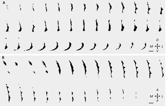
Figure 3 .

The claustrum in coronal and axial views. Sequential slices of the automatically segmented right claustrum in 1 subject. (A) Coronal slices from posterior to anterior. (B) Axial slices from ventral to dorsal. Scale bar: 10 mm. See also Supplementary Video 1 for a 3D representation. Key: M: medial, L: lateral; V: ventral; D: dorsal; P: posterior; A: anterior.
