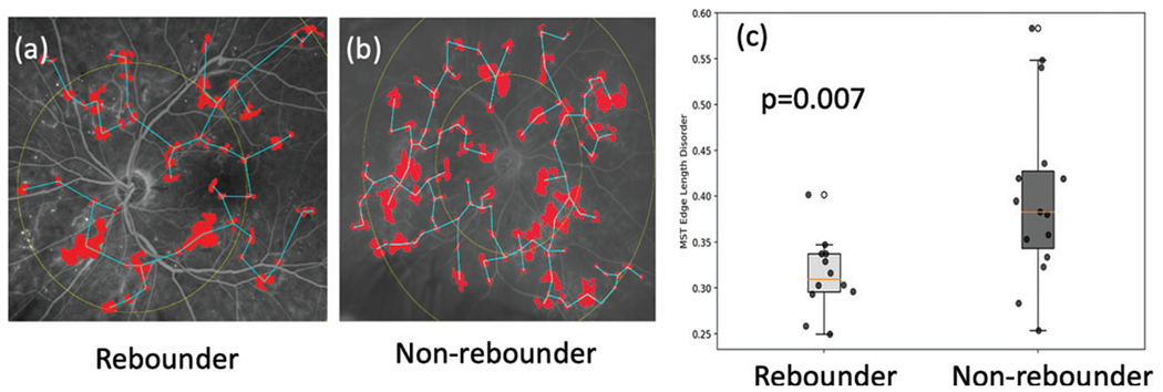
Figure 2.

Leakage feature extraction. (A) and (B) are example baseline FA images of a non-rebounder and a rebounder, respectively. Their corresponding leakage patches are highlighted in red, and the minimum spanning tree edges in blue. Centroids of leakage patches are used as nodes and vectors connecting them are edges. Weights are the length of the edges. (C) Box and whisker plot on the left corresponds to the MST edge length disorder values from the rebounders, and the one on the right corresponds to the MST edge length disorder from the non-rebounders. FA, fluorescein angiography; MST, minimum spanning tree.
