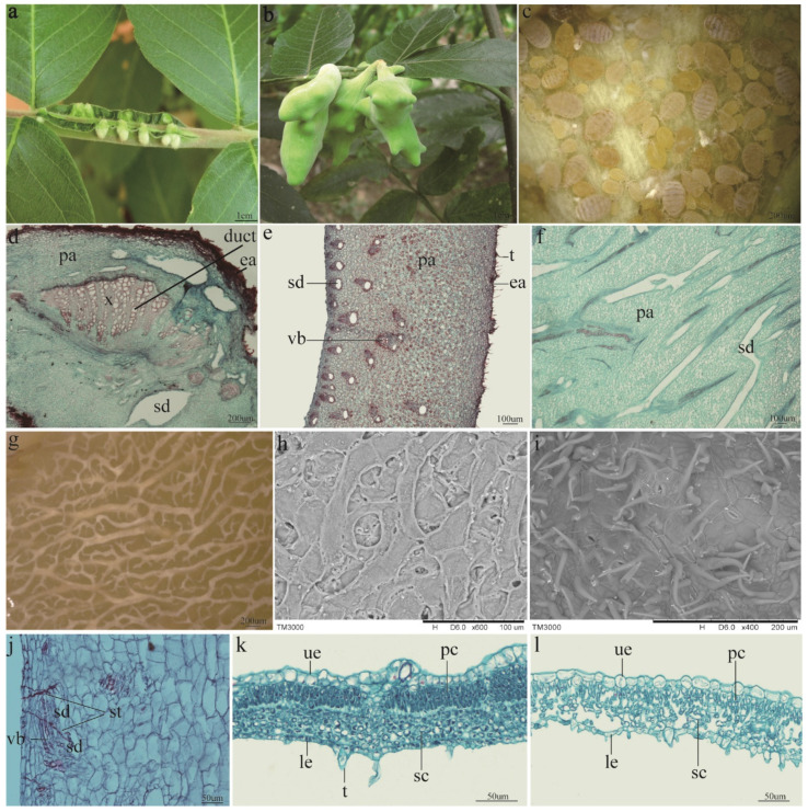
Figure 1.
Localization, morphology, and anatomical structure of the horned gall. (a) Young horned galls clustered on rachis wings. (b) Mature galls connected with rachis leaf wing via a specialized stalk. (c) Aphids feeding on inner wall. (d) Cross-section of the stalk. Many ducts are present in the expanded xylem. (e) Crosscutting of the gall wall. The wall comprises parenchyma cells and contains many vascular bundles that are joined to the schizogenous ducts. (f) Plane surface of the horned gall; long schizogenous ducts are present. (g) The inner surface of the horned gall treated by NaOH. Vast, branched schizogenous ducts are present in the inner wall. (h) A scanning electron microscope (SEM) image of the inner surface. The inner wall was rough and characterized by holes. (i) The outer surface of the horned gall in the SEM image. Stoma and tomentum are present in the outer wall. (j) The stylophores of the aphids. Stylophores gathered in the vascular bundle of the horned gall. (k) The cross-section of the young R. chinensis leaf. The palisade and spongy cells are closely arranged. (l) The cross-section of the mature R. chinensis leaf. The cells, particularly the spongy tissues, are loose and porous. ea = epidermis-air, x = xylem, pa = parenchyma, sd = schizogenous duct, vb = vascular bundle, t = tomentum, st = stylophore, el = epidermis-lumen, pc = palisade cell, sc = spongy cell, le = lower epithelial cell, ue = upper epithelial cell.

