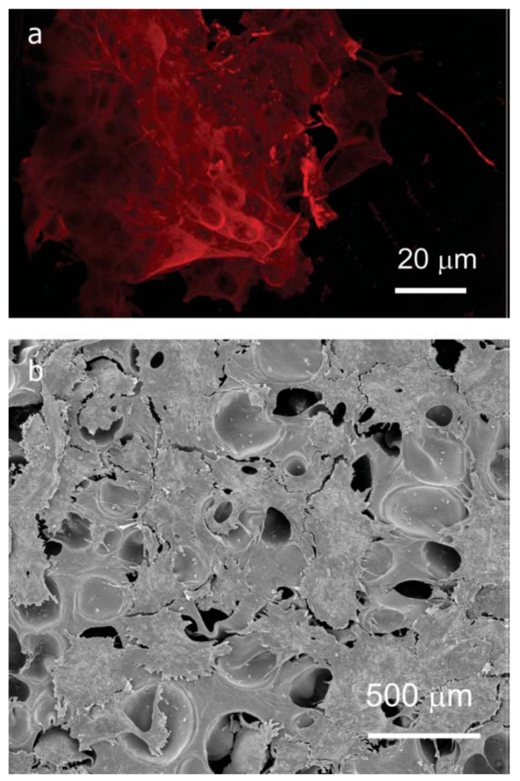
Figure 20.
(a) Fluorescent microscopy image of neuroblastoma (human neuron-like, SH-SY5Y) cells cultured on foamed 30/70 SIS/THFMA blends. Note the intense red staining of the neuronal cytoskeletal protein b-III tubulin in the cluster of cells shown here. (b) Electron micrograph of ovine meniscal chondrocytes cultured on foamed 30/70 SIS/THFMA blends [297].

