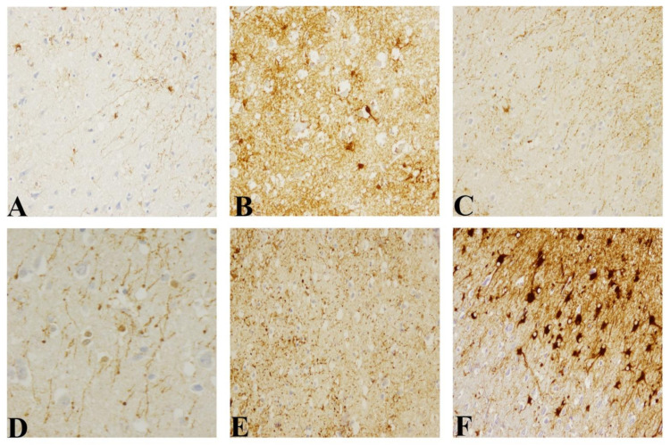
Figure 5.
In comparison with that in healthy control, (A) morphological changes of interlaminar astroglia in CJD cases are illustrated (GFAP antibody). (B) Replacement by typical stellate astroglial cells with increase of astrogliosis. (C) Increase of number of terminal masses of interlaminar astroglia. (D) Increase of diameter of terminal masses. (E) Disruption in I–III frontal cortex. (F) Gemistocytic phenotype in panencephalopathic CJD (Magnification: 200×, except for (D) 400×).

