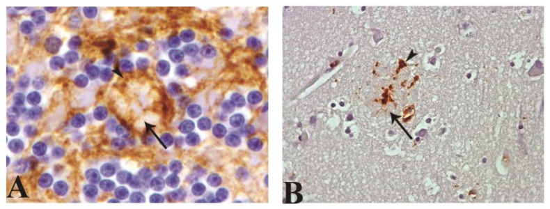
Figure 6.
Illustration of glial cells associated to protein deposits by astroglial (GFAP antibody; arrowhead) and microglial (CD68 and MHCII antibodies; arrowhead) immunostaining, respectively. (A) Astrocytic process associated with Kuru plaque (arrow) in granular layer of cerebellum. (B) Microglial process associated to protein aggregation or clustered morphology associated with protein deposit (arrow; magnification: 40×).

