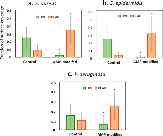
Figure 5.

Quantitative analysis of the live and dead fractions of (a) S. aureus, (b) S. epidermidis, and (c) P. aeruginosa colonization on hydrogel surfaces with and without AMP attachment. Values were obtained from analyzing fluorescent images using ImageJ software. Values are mean ± SD, triplicate samples were used, and three independent experiments were performed (n = 3), *p < 0.05 compared to control groups (hydrogels with no AMP).
