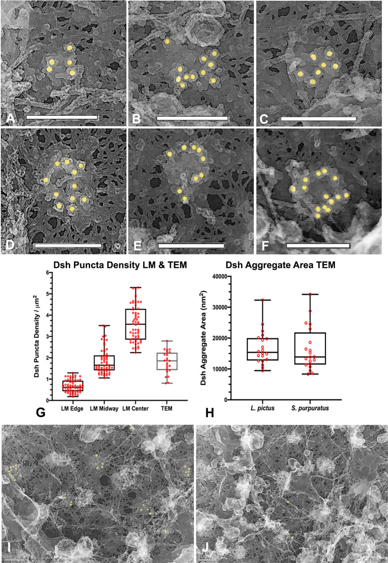
Fig 3. Platinum replica TEM demonstrates that Dsh puncta correspond to patches of aggregates of pedestal-like structures.
(A-F) A gallery of high magnification images of Dsh labeled structures demonstrates that they consist of aggregates of pedestal-like structures in a variety of groupings. The top of each pedestal is labeled with a single colloidal gold particle suggesting that this area may correspond to the location of the C terminus of the Dsh protein. (G) The density of the Dsh puncta in TEM images from 3 separate cortices over three experiments (grey box) falls in the range of densities seen in the immunofluorescence images (black boxes reproduced from graph in Fig 1J). (H) The area of the Dsh aggregates from five cortices each from two separate experiments shows no statistically significant difference between the two species. (I,J) Comparison of regions of the same cortex in which Dsh labeling is present (I) and is not present (J) shows that the patches associated with the Dsh labeling are not visible in the unlabeled regions, suggesting that these structures are specific to the Dsh puncta. L. pictus cortices = A,B,D,E; S. purpuratus cortices = C,F,I,J. Scale bars = 200 nm.

