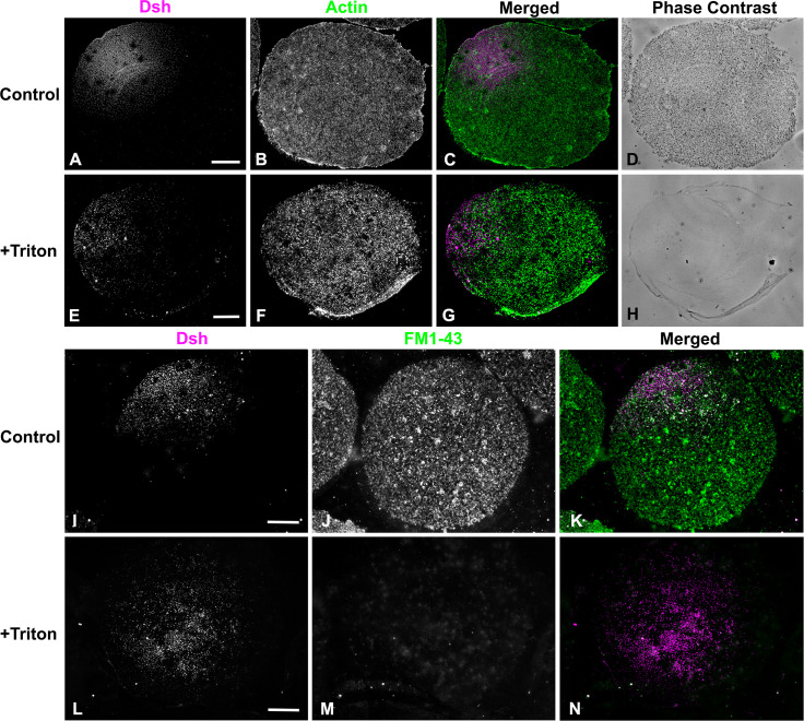
Fig 4. The Dsh VCD array is resistant to Triton detergent extraction.
(A-H) Control unfertilized egg cortex from L. pictus (A-D) stained for Dsh (magenta) and actin (green) showing Dsh array and cortical granules in phase contrast. The Triton extracted cortex (E-H) demonstrates the persistence of the Dsh array following detergent extraction despite the loss of cortical granules seen in phase contrast. (I-N) Membrane staining with the fixable dye FM1-43 (green) shows that in control unfertilized egg cortices (I-K) that the Dsh (magenta) array is present along with a variety of membranous structures. In detergent extracted cortices (L-N) the Dsh array is still present even though specific membrane staining is lost. Scale bars = 10 μm.

