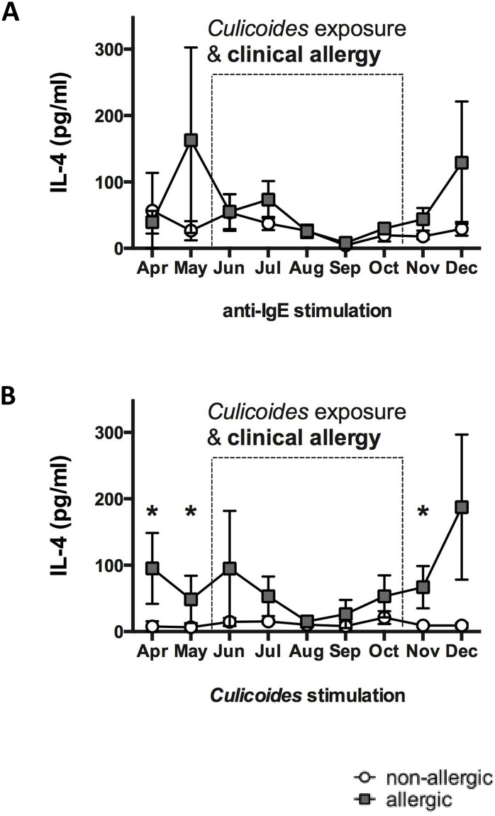
Fig 1. Anti-IgE and Culicoides (Cul) stimulation induced IL-4 secretion in PBMC of allergic and non-allergic horses.
Blood samples were obtained monthly from allergic (n = 8) and non-allergic horses (n = 8) from April to December. The dotted box indicates the period of environmental exposure to Cul when horses with CH showed clinical signs of allergy. PBMC were stimulated in vitro with different stimuli, cell culture supernatants were harvested after 24 hours of incubation, and IL-4 was measured in the supernatants using a fluorescent bead-based IL-4 assay. IL-4 secretion from PBMC after stimulation with A) anti-IgE, clone 134, which crosslinks IgE on the cell surface, and B) Cul extract. Graphs represent means with standard errors. Allergic and non-allergic groups were compared using non-parametric Mann Whitney tests. * p<0.05.

