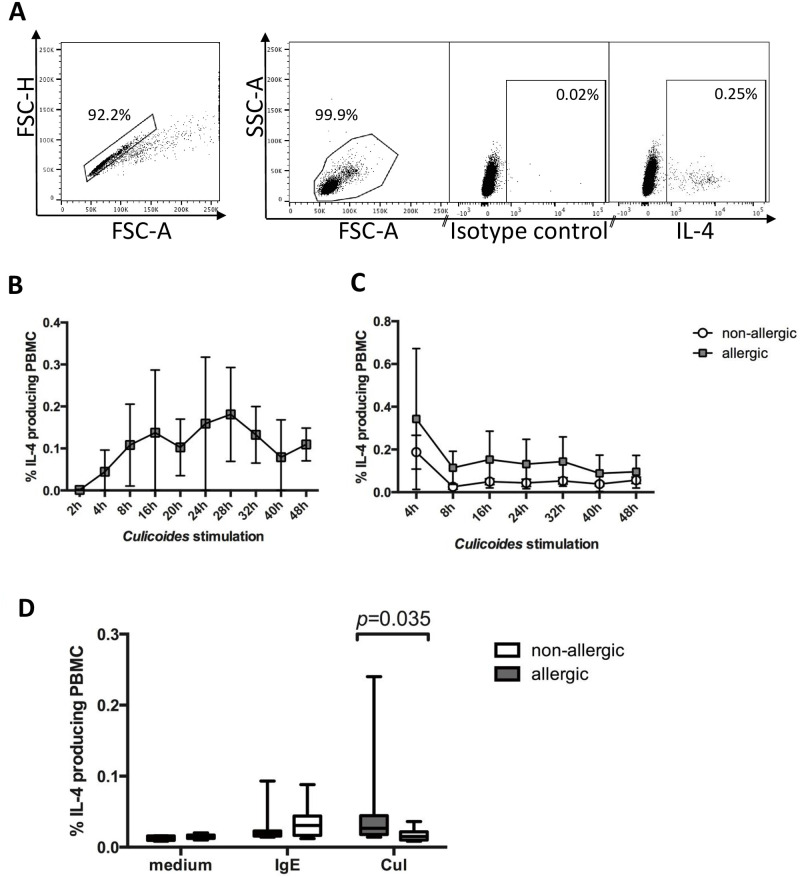
Fig 2. Flow cytometric analysis of IL-4+ cells after stimulation of PBMC from allergic and non-allergic horses with anti-IgE or Cul extract.
PBMC were stimulated in vitro with crosslinking anti-IgE mAb 134 or Cul in the presence of the secretion blocker Brefeldin A. After incubation, the cells were fixed, stained for intracellular IL-4 production, and analyzed by flow cytometry. A) Gates were set for doublet exclusion (FSC-H/FSC-A) and on PMBC by forward and side scatter characteristics (FSC-A/SSC-A). PBMC were then analyzed for IL-4 expression in comparison to the isotype control. B) PBMC from two allergic horses were stimulated with Cul in the presence of the secretion blocker Brefeldin A for different times between 2–48 hours. Cells were fixed after incubation and stained for intracellular IL-4. C) PBMC from allergic (n = 5) and non-allergic horses (n = 5) were stimulated for 4–48 hours with Cul and Brefeldin A was only added for the last four hours of incubation. D) Percentages of IL-4+ PBMC from horses with CH (n = 8) and non-allergic controls (n = 8) were isolated in August, stimulated for four hours with anti-IgE 134 or Cul or kept in medium as control.

