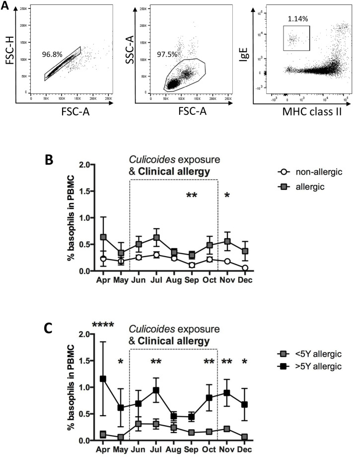
Fig 3. Basophils in the peripheral blood of allergic and non-allergic horses were compared by flow cytometric analysis.
PBMC from sixteen Icelandic horses were analyzed once a month with cell surface staining for IgE and MHC class II. A) For analysis of basophils, cells were gated for doublet exclusion (left) and by forward (FSC) and side scatter (SSC) characteristics (middle). MHCII expression and IgE on gated PBMC was used to gate on basophils (IgE+/MHCIIlow cells; right). B) Percentages of basophils were compared over time between allergic (n = 8) and non-allergic (n = 8) horses and C) in horses with allergy for >5 years (n = 4) and those with allergy <5 years (n = 4). The dotted box marks the period of exposure to Cul and clinical signs in the allergic horses. Data represent means and standard errors. Horse groups are compared by a non-parametric Mann Whitney Test. * p<0.05; ** p<0.01 **** p < 0.0001.

