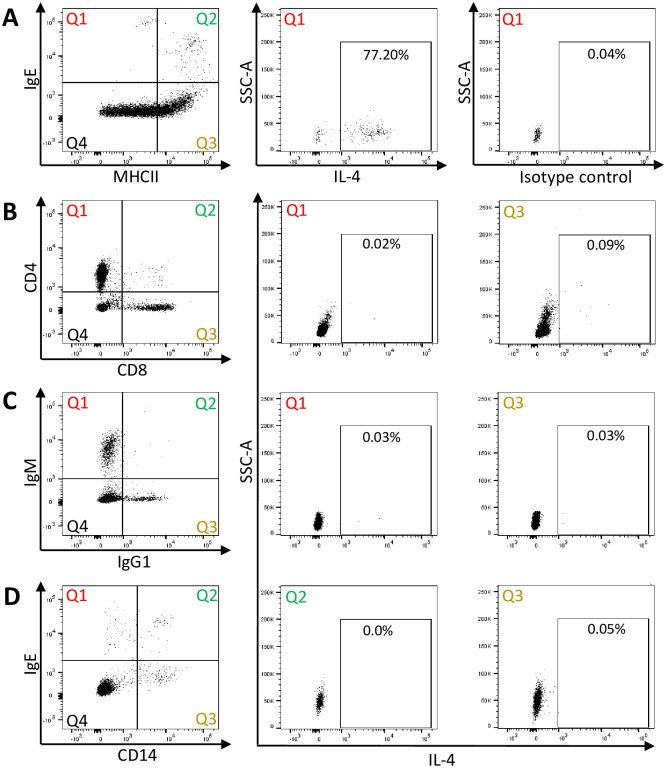
Fig 4. Phenotyping of IL-4+ peripheral blood cells after stimulation with Cul extract in vitro.
PBMC were stimulated with Cul in presence of secretion blocker Brefeldin A for 4 hours. For flow cytometry analysis, cells were fixed and stained for intracellular IL-4 and subsequently for extracellular surface proteins: A) IgE and MHCII expression of PBMC to identify basophils in Quadrant (Q)1 (left), IL-4 production by IgE+/MHCIIlow basophils (middle) and an intracellular isotype control staining (right); B) CD4 and CD8 expression to identify T cells (left), IL-4 production by CD4+ T cells in Q1 (middle), and by CD8+ T cells in Q3 (right); C) IgM and IgG expression to identify B cells (left), IL-4 production by IgM+ B cells (middle), and by IgG1+ cells (right); D) IgE and CD14 expression to identify monocytes (left), IL-4 production by IgE+/CD14+ IgE-binding monocytes (middle), and by IgE-/CD14+ monocytes (right). The IL-4+ cells give the percentages from the respective quadrant analysis. Representative flow cytometry plots from one horse in August are shown. The analysis was performed on all 16 horses.

