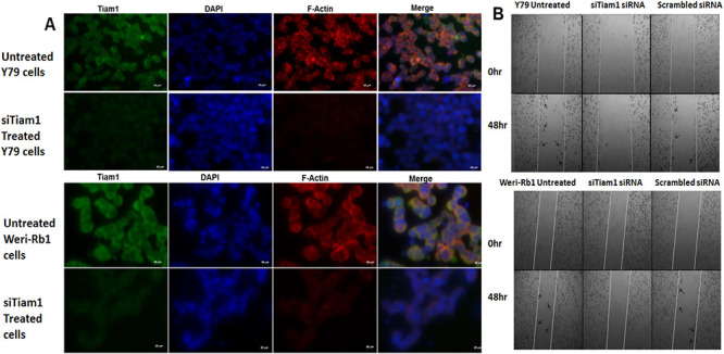
Fig 5. F-actin staining and invasion of Tiam1 deficient RB cell lines.

A. Tiam1 silenced Y79 cells and Weri-Rb1 cells were fixed, immunofluorescently labeled for Tiam1, nucleus stained with DAPI, stained with phalloidin and images were taken at 40X in ten fields. Bar represents 20 μm. B. Phase contrast microscope images of wound healing assay showing the cell migration pattern in Tiam1 deficient retinoblastoma cell lines at 0 hr and 48 hrs post silencing. Tiam1 knockdown cells were unable to migrate whereas the untransfected and control cells showed increased migration, migrated cells were indicated with arrows. The images were acquired using AxioObserver microscope at 5× objective with 1× optovar.
