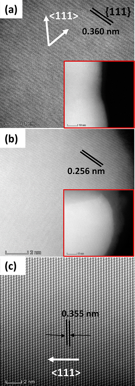
Figure 3.

STEM analysis of Ge1–xSnx nanowires. (a) STEM image for Ge1–xSnx nanowire with high (∼20 atom %) Sn content. The low resolution of the image is associated with the deformation of the Ge1–xSnx nanowire by the electron beam. The inset shows low-resolution STEM of the corresponding nanowire. (b) STEM image of the spherical tip after the nanowire growth shows the formation of a predominantly Sn rich part at the tip. (c) High resolution STEM image of Ge1–xSnx nanowire with around 10 atom % Sn shows high crystal quality and ⟨111⟩ growth direction.
