Figure 1.
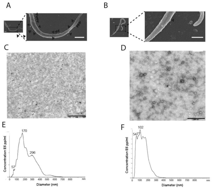
Purification of the extracellular vesicles (EVs) of epimastigotes (E) and EVs of tissue-culture cell-derived trypomastigotes (TCT) of T. cruzi (Pan4 strain, DTU I). Scanning electron microscopy (SEM) of E (A) and TCT (B) employed in this study (scale bar: 1 µm). (C) Transmission electron microscopy (TEM) of purified EVs of E (scale bar: 1 µm). (D) TEM of purified EVs of TCT (scale bar: 500 nm). (E) Nanoparticle tracking analysis (NTA) size distribution of purified EVs of E. (F) NTA size distribution of purified EVs of TCT. Representative figures and graphs of 7 different replicates are shown.

