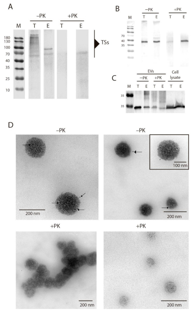
Figure 4.

Location of TS proteins on the surface of EVs of T. cruzi. (A) Western blot of EVs of E and EVs of TCT treated/untreated (+PK/−PK, respectively) with proteinase K and incubated with anti-TS antibodies. All lanes were loaded with an equal amount of protein (30 µg). (B) Western blot of EVs of E and EVs of TCT treated/untreated (+PK/−PK, respectively) with proteinase K and incubated with anti-cruzipain antibodies. All lanes were loaded with an equal amount of protein (30 µg). (C) Western blot of EVs of E and EVs of TCT treated/untreated with proteinase K and whole lysates of TCT and E incubated with anti-calmodulin antibodies. All lanes were loaded with equal amount of protein (30 µg). (D) Immunogold labeling of TS proteins in EVs of TCT treated/untreated with proteinase K. Black arrows indicate the gold particles and thus TS location.
