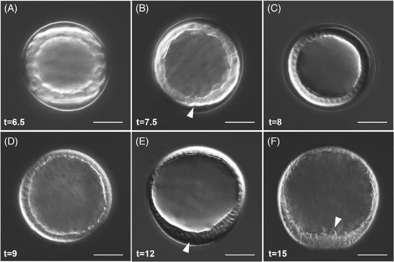
FIGURE 2.
Blastula stages, hatching, and early ingression of PMCs in L. pictus. A, Early blastula stage. B, Embryos begin to demonstrate cell shape changes, and the small micromeres (white arrow) are visible at the vegetal pole. C, Blastula prehatching. D, Hatched blastula. E, Mesenchyme blastula. White arrow points at the thickening at the vegetal plate. F, White arrow points at PMCs ingressing into the blastocoel, which are visible at the vegetal pole. For all panels, scale = 50 μm. Times listed in hours postfertilization (hpf)

