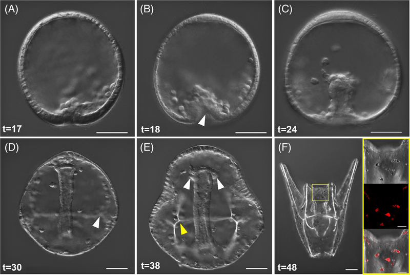
FIGURE 3.
Gastrulation, SMC differentiation, and skeletal rod formation in L. pictus. A, First signs of invagination of the vegetal pole are apparent after ingression of PMCs. B, Primary gastrulation completes and the vegetal pole is turned inward as indicated by the white arrow. A population of SMCs ingress into the blastocoel and the PMCs begin to arrange around the developing archenteron. C, During mid-gastrulation the archenteron moves through the blastocoel and pigment cell precursors will migrate through the blastocoel to embed into the ectoderm during mid-late gastrulation. D, Late gastrulae have clear arrangement of PMCs and triradiate spicules which will further develop into the larval skeleton (white arrow). The archenteron has nearly reached the oral side of the animal. E, Evidence of the forming coelomic pouches (white arrows) on either side of the archenteron preclude the fusion of the mouth with the oral ectoderm during the prism larval stage, and the arms begin to bud out from the larval body as the skeletal supports (yellow arrow) are further elaborated. F, Composite stack of early pluteus larva in abanal view. The gut has differentiated into three parts. A yellow box surrounds the region of the larva shown in the inset. Inset shows DIC, fluorescence, and overlay of pigment cells which contain the autofluorescent pigment echinochrome A and are embedded into the ectoderm of the larva. For all panels, scale = 50 μm. Inset panels scale = 20 μm. Times listed in hours post-fertilization (hpf)

