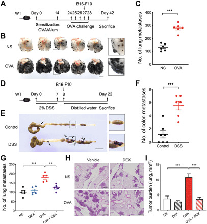
Fig. 1. Airway inflammation and colonic inflammation promotes metastasis.

(A) Schematic timeline representing the merger of an established model of allergic airway inflammation and the metastasis of circulating B16-F10 cells to the lung. (B) Representative photograph of pulmonary metastatic foci on day 42. Scale bar, 5 mm. (C) The number of experimental pulmonary metastases. (D) Schematic timeline representing the merger of an established model of colitis and the metastasis of abdominal B16-F10 cells to the colon. (E) Representative photograph of colonic metastatic foci produced after injection of B16-F10 cells on day 22. Scale bar, 1 cm. (F) The number of colon metastases. (G) The number of experimental pulmonary metastases from mice treated with NS, dexamethasone (DEX), OVA, and OVA and DEX (OVA + DEX), respectively. (H) Representative lung hematoxylin and eosin section of metastases. Scale bars, 500 μm. (I) Quantification of the tumor burden per lung (mm2) in (H). (C, F, G, and I) n = 5 to 7 mice per group. Statistical analyses were performed by Student’s t test (C and F) and one-way ANOVA (G and I). Data are presented as mean ± SEM from one representative experiment of three independent experiments. **P < 0.01; ***P < 0.001.
