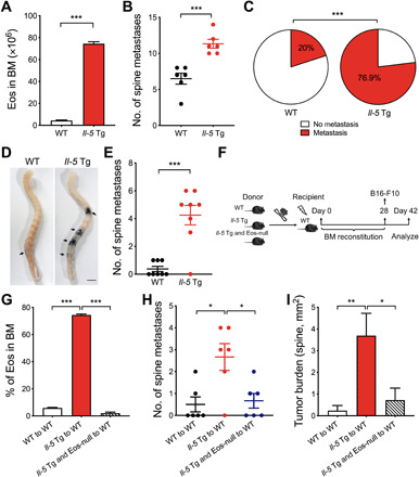
Fig. 2. Eosinophilia mediates enhanced bone metastasis in Il-5 Tg mice.

(A) The number of eosinophils in BM of WT and Il-5 Tg mice, n = 4 mice per group. (B) Comparison of metastasis foci of spines on day 14. WT and Il-5 Tg mice were injected with B16-F10 cells via caudal arteries. (C) The proportion of metastasis in the spine from mice injected with B16-F10 intravenously (WT, n = 20; Il-5 Tg, n = 26). (D) Representative photograph of metastatic foci in the spine of WT and Il-5 Tg mice injected with B16-F10 intravenously on day 14. (E) The number of the spine metastasis in (D). (F) Schematic representation of BM transplantation followed by establishing a metastasis model. (G) Frequencies of eosinophils in BM of WT mice that have received donor BM transplants from WT, Il-5 Tg, or Il-5 Tg and Eos-null mice 28 days ago. (H) Comparison of the metastatic foci in the spine on day 42. (I) Quantification of the tumor burden per area (mm2) in the spine on day 42. (B, E, and G to I) n = 6 to 8 mice per group. Statistical analyses were performed by Student’s t test (A, B, and E), one-way ANOVA (G, H, and I), and Fisher’s exact test (C). Data (mean ± SEM) are from one representative experiment of three independent experiments. *P < 0.05, **P < 0.01, and ***P < 0.001.
