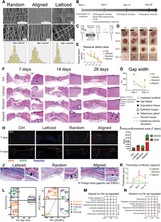
Fig. 1. Evaluation of the healing of rat skin wounds implanted with three types of electrospun scaffolds.

(A) Surface topography of random, aligned, and latticed electrospun membranes. (B) Workflow for evaluating rat skin wound healing. PCR, polymerase chain reaction. (C) Surgical processes for the rat skin excisional wound model. (D and E) Residual wound area at 3, 5, 7, and 14 days. Photo credit: Chen Hu, Sichuan University. (F) Histological view of the Control (Ctrl) and groups implanted with three types of scaffolds. Regenerated epithelium showed stratified structure at 14 days. (G) Semiquantitative evaluation of gap width. (H) Immunofluorescent staining using Krt5 (red) and Krt10 (green). (I) Semiquantitative evaluation of the fluorescent area. (J) Evaluation of foreign body reaction around the biomaterials. Foreign body gigantic cells lined up on the random and latticed membranes, whereas fewer of them appeared on aligned membranes. (K) Thickness of fibrotic capsules around random, aligned, and latticed membranes. (L to N) Principle components (PC) analysis and GO analysis showing immunomodulatory effects for aligned and random scaffolds. ****P < 0.0001, **P < 0.01, and *P < 0.05 by analysis of variance (ANOVA) for data in (E), (G), and (K).
