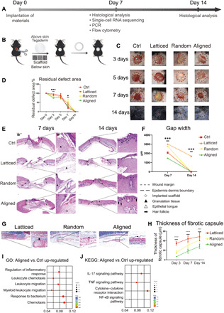
Fig. 2. Evaluation of the healing of mouse skin wounds implanted with three types of electrospun scaffolds.

(A) Workflow for evaluating mouse skin wound healing. (B) Surgical processes for mouse skin excisional wound model. (C and D) Residual wound area at 3, 5, 7, and 14 days. Photo credit: Chenbing Wang, Sichuan University. (E) Histological analysis of Ctrl and other groups implanted with three types of scaffolds. (F) Semiquantitative evaluation of gap width. (G) Evaluation of foreign body reaction around biomaterials. (H) Thickness of fibrotic capsules around random, aligned, and latticed membranes. (I and J) GO and KEGG analysis, respectively, showing immunomodulatory effects for aligned scaffolds. ****P < 0.0001, ***P < 0.001, and *P < 0.05 by ANOVA for data in (D), (F), and (H). NF-κB, nuclear factor κB; TNF, tumor necrosis factor.
