
Fig. 4. Validation of models and immunofluorescence imaging of lung tissue.
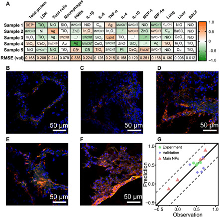
(A) Prediction errors of the validation sets. DEPa, diesel engine particles; DWCNTb, double-wall carbon nanotubes; SWCNTb, single-wall carbon nanotubes; …d, SWGe-imogolite; CBe, carbon black; …f, cellulose nanocrystals; …g, QD-CdSe-ZnS; …h, Rosette nanotubes. (B) Immunofluorescence imaging of control. (C) F-MWCNTs. (D) S-MWCNTs. (E) M-MWCNTs. (F) L-MWCNTs. (G) Validation of models using IL-1β fluorescence intensity. Red channel, nuclear factor κB (NF-κB) p65; green channel, IL-1β; and blue channel, 4′,6-diamidino-2-phenylindole (DAPI).