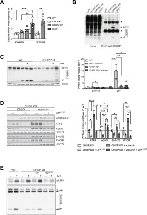
Fig. 6. CHOP interacts with C/EBPb protein to repress the ATF4 activation.

(A) Relative Gadd34 transcript levels at P17 (±2) WT, CHOP KO, DARS2 KO, and DKO animals, as well as in 6-week-old WT and DARS2 KO mice. Bars represent means ± SD, samples were normalized to WT mice of the respective age (P17: one-way ANOVA, *P < 0.05, **P < 0.01, and ***P < 0.001; 6 weeks: unpaired Student’s t test) (n = 4). (B) Coimmunoprecipitation (co-IP) of CHOP from WT, CHOP KO, DARS2 KO, and DKOL hearts. The CHOP and C/EBPβ interaction was monitored with Western blotting using an antibody against C/EBPβ. One percent of the input fractions was used as loading controls. Asterisks indicate the immunoglobulin G heavy and light chains. (C) Western blot analysis (left) and quantification (right) of the three CEBPβ isoforms LAP1, LAP2, and LIP in CHOP KO MEFs treated for 48 hours with actinonin along with the respective control (n = 3). (D) Western blot analysis (left) and quantification (right) of steady-state levels of ISR markers in actinonin-treated (48 hours) CHOP KO MEFs expressing the CEBPβ LIPL120T mutant variant along with the respective controls (n = 3). (E) Western blot analysis of the ATF4 and three CEBPβ isoforms in actinonin-treated (48 hours) CHOP KO MEFs expressing the CEBPβ LIP WT and CEBPβ LIPL120T mutant variant along with the WT cells and respective controls (n = 4). (C to E) Antibodies used were raised against proteins indicated in the panels. HSC70 was used as a loading control. (A, C, and D) Bars represent means ± SD (MANOVA followed by one-way ANOVA and Tukey’s multiple comparisons test, *P < 0.05, **P < 0.01, ***P < 0.001, and ****P < 0.0001) (n = 3).
