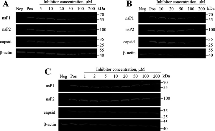
Figure 7.
Western blot analysis of proteins from CHIKV-infected cells that were treated using compounds D160a (R,R,S-stereoisomer) (A), D161 (an equimolecular mixture of four possible stereoisomers) (B), and D160d (S,S,S-stereoisomer) (C). BHK-21 cells infected with CHIKV-NanoLuc (MOI 10) were treated with increasing concentrations of the inhibitor. Cell lysates were collected 6 h post infection; proteins were separated using SDS-PAGE, transferred onto the PVDF membrane, and detected using indicated antibodies. β-Actin was detected as the loading control. Names of the proteins are indicated on the left; molecular masses of marker bands are indicated on the right. Neg—mock-infected BHK-21 cells treated with 1% DMSO; Pos—CHIKV-infected BHK-21 cells treated with 1% DMSO (no inhibitor).

