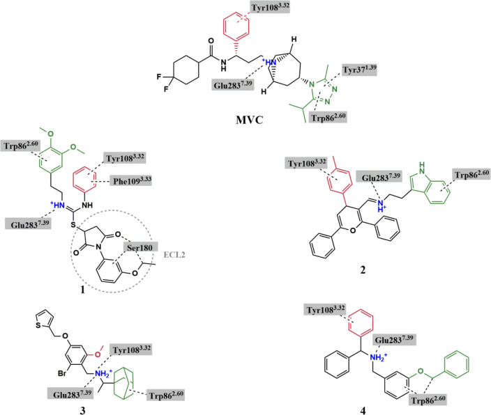
Figure 3.
Chemical structures of hit compounds showing the basic nitrogen (blue) and functional groups fitting in TMS1 (green) and the hydrophobic subpocket of TMS2 (red). Interactions between the ligands and specific residues derived from the CCR5 X-ray structure (PDB code: 4MBS) are depicted by dotted lines. Structure of MVC is added for comparison.

